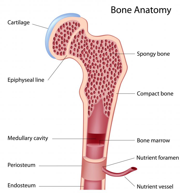
Histology Of Compact Bone Diagram : Structure of compact bone your skills & rank.. Use the venn diagram to compare and contrast compact bone and spongy bone. Haversian canal, lacuna, circumferential lamella, interstitial lamellae, osteocyte, canaliculi, matrix, osteon 2. Good, here in this part, i am going to describe the structure of compact bone. As seen in the image below, compact bone forms the cortex, or hard outer shell of most bones in the body. The most robust aspect of this unit is the underlying bony architecture.
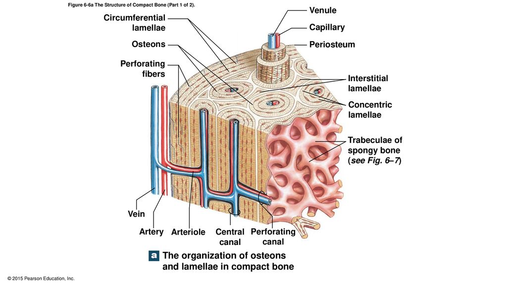
Diagramme schnell und einfach erstellen. Immature (streamer) bone due to haphazard (random) arrangement of collagen fibers, found during growth, healing, repair, infections or in some neoplasms. Compact bone is the denser, stronger of the two types of osseous tissue (figure 6.3.6). The diagram above shows a longitudinal view of an osteon. They are sketches from selected slides used in class from the teaching slide set.
Osteon Wikiwand from upload.wikimedia.org Good, here in this part, i am going to describe the structure of compact bone. Compact bone, or cortical bone, mainly serves a mechanical function. The differences between compact and spongy bone are best explored via their histology. The diagram above shows a longitudinal view of an osteon. Outside all of bone, is a connective tissue sheath called the periosteum (see below photograph). It is thick and dense. There are two types of mature bone: Growth plate, made of cartilage, gradually ossifies.
Compact bone, also called cortical bone, dense bone in which the bony matrix is solidly filled with organic ground substance and inorganic salts, leaving only tiny spaces (lacunae) that contain the osteocytes, or bone cells.compact bone makes up 80 percent of the human skeleton;
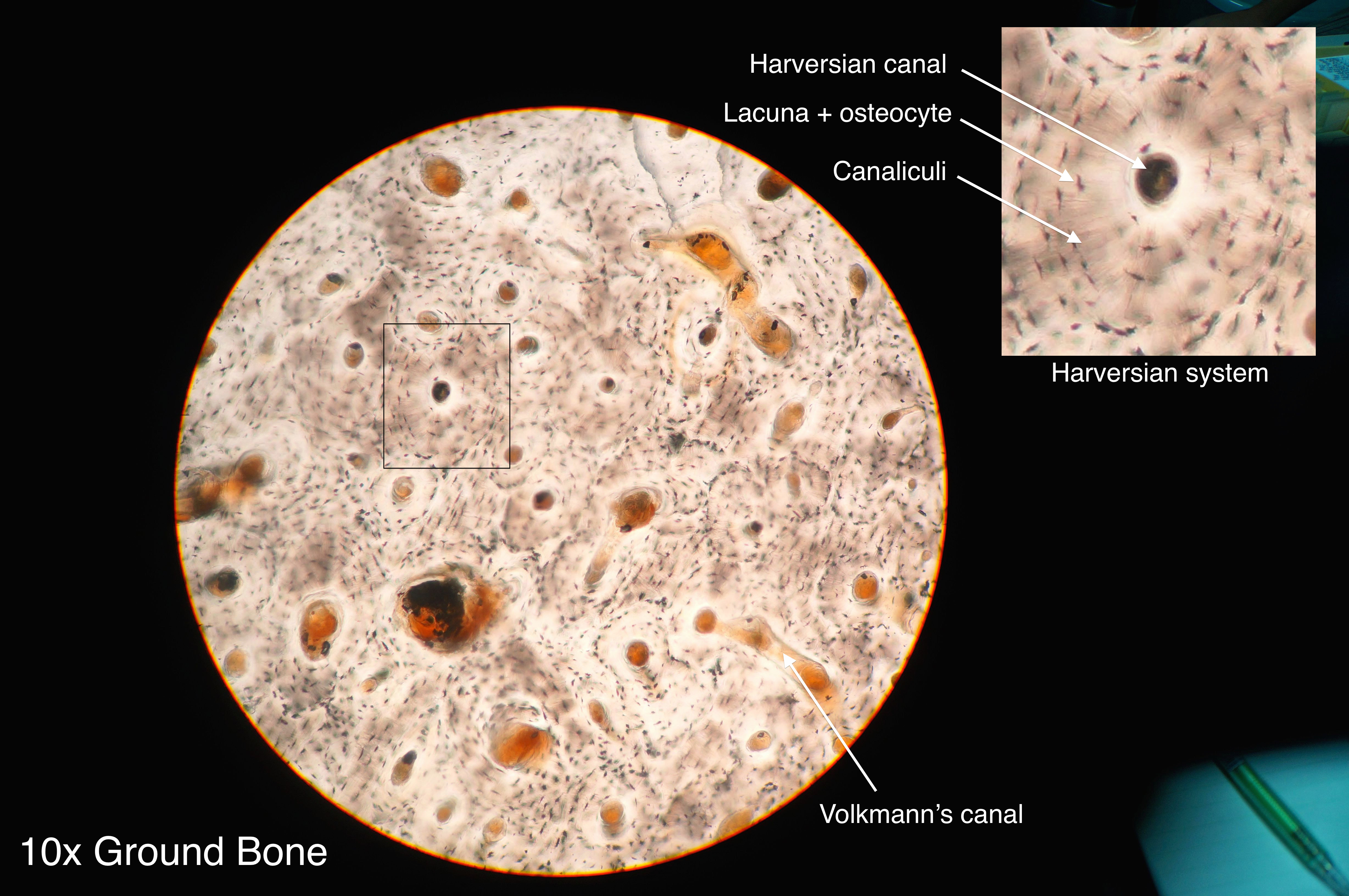
In compact bone, you will find the three bone. A thin layer of compact bone also covers the epiphyses of long bones. The inner space is lined by osteoblasts and osteoclasts (called the endosteum). Each osteon looks like a ring with a light spot in the center. This short article will describe the spongy bone histology and labeled diagram and real slide pictures. Compact bone is the denser, stronger of the two types of bone tissue ( (figure) ). In long bones, spongy bone forms the interior of the epiphyses; So, this is a slide of compact bone histology. This is an online quiz called structure of compact bone. Begin by identifying the concentric rings of lamellar bone that surround a haversian canal. Bones are the organs of the skeletal system; As seen in the image below, compact bone forms the cortex, or hard outer shell of most bones in the body. Outside all of bone, is a connective tissue sheath called the periosteum (see below photograph).
Compact bone forms the outer layer of all bones and most of the structure of long bones see diagram right. A thin layer of compact bone also covers the epiphyses of long bones. Good, here in this part, i am going to describe the structure of compact bone. This type of bone is located between layers of compact bone and is thin and porous. Histology fundamentals bone structure amp types draw it to know it there are three types of cells that contribute to bone homeostasis.
What Is Compact Bone With Pictures from images.infobloom.com The drawings of histology images were originally designed to complement the histology component of the first year medical course run prior to 2004. Start studying histology of compact and spongy bone. As seen in the image below, compact bone forms the cortex, or hard outer shell of most bones in the body. In long bones, spongy bone forms the interior of the epiphyses; They are sketches from selected slides used in class from the teaching slide set. Use the venn diagram to compare and contrast compact bone and spongy bone. Each osteon looks like a ring with a light spot in the center. The first bone formed at any site is woven (or primary) bone, but this is soon replaced by lamellar bone.
This type of bone is located between layers of compact bone and is thin and porous.
Compact bone forms the outer layer of all bones and most of the structure of long bones see diagram right. (b) in this micrograph of the osteon, you can clearly see the concentric lamellae and central canals. The strength, shape and stability of the human body are dependent on the musculoskeletal system. There is a printable worksheet available for download here so you can take the quiz with pen and paper. Mineralized bone is stained black and soft tissues are stained light blue. Cavity within the shaft of the long bones filled with bone marrow. Spongy bone spongy bone is the tissue that makes up the interior of bones; The first bone formed at any site is woven (or primary) bone, but this is soon replaced by lamellar bone. Haversian canal, lacuna, circumferential lamella, interstitial lamellae, osteocyte, canaliculi, matrix, osteon 2. In long bones, spongy bone forms the interior of the epiphyses; Each osteon looks like a ring with a light spot in the center. It can be found under the periosteum and in the diaphyses of long bones, where it provides support and protection. Hey there, welcome back again to anatomy learner and thank you so much for getting into this article.
Thin layer of reticular ct lining internal marrow cavity. Highlighted with polarized light or reticulin stain. Mineralized bone is stained black and soft tissues are stained light blue. Begin by identifying the concentric rings of lamellar bone that surround a haversian canal. Trabecular bone, also known as cancellous bone or spongy bone, mainly serves a metabolic function.
6 4 Compact Bone And Spongy Bone Ppt Download from slideplayer.com Highlighted with polarized light or reticulin stain. Bones are the organs of the skeletal system; Hard, dense bone tissue that is beneath the outer membrane of a bone. The diaphysis (shaft) consists of compact bone surrounding the central marrow cavity. Use the venn diagram to compare and contrast compact bone and spongy bone. These labelled diagrams should closely follow the current science courses in histology, anatomy and Cavity within the shaft of the long bones filled with bone marrow. Compact bone is the tissue that forms the surface of bones.
The diagram above shows a longitudinal view of an osteon.
Diagramme schnell und einfach erstellen. The diaphysis (shaft) consists of compact bone surrounding the central marrow cavity. Some, mostly older, compact bone is remodelled to form these haversian systems (or osteons). It can be found under the periosteum and in the diaphyses of long bones, where it provides support and protection. Compact bone is the denser, stronger of the two types of osseous tissue (figure 6.3.6). Spongy bone spongy bone is the tissue that makes up the interior of bones; Hard, dense bone tissue that is beneath the outer membrane of a bone. Compact bone definition compact bone, also known as cortical bone, is a denser material used to create much of the hard structure of the skeleton. This short article will describe the spongy bone histology and labeled diagram and real slide pictures. There are two types of mature bone: The light spot is a canal that carries a blood vessel and a nerve fiber. Highlighted with polarized light or reticulin stain. This is an online quiz called structure of compact bone.
Structure of compact bone your skills & rank compact bone diagram. The most robust aspect of this unit is the underlying bony architecture.


0 Komentar